In an effort to improve outcomes for patients diagnosed with gliomas—tumors that account for about three in ten of all tumors that start in the brain—members of the Neurosurgery team at the UTMB Health Clear Lake Hospital are now conducting
fluorescence-guided surgery to remove these masses.
.jpg?sfvrsn=94037056_1)
This approach to brain surgery has been studied for years by UTMB neurosurgeon Dr. Pablo Valdes,
who dedicated much of his PhD work to this science. Patients ingest a medicated drink that contains 5-aminolevulinic acid (5-ala) two to four hours before the start of anesthesia for their surgery.
The medication then leads to an accumulation of a fluorescent marker, sometimes called a fluorophore, within the body. When viewed under a special light, the marker will emit violet-blue hues from the healthy parts of the brain anatomy, while the
abnormal tumor will display reddish-pink.
The contrast helps surgeons more easily identify what needs to be removed, versus what needs to stay.
“The medicine helps us color tag the tumor and conduct color-coded surgery,” said Dr. Valdes, adding that the practice has been in use at the UTMB Galveston Campus for some time, before the recent expansion to the services offered in the
Bay Area.
While the description sounds simple, it’s a technology that’s allowing doctors to get within millimeters of critical areas when removing the potentially malignant threats.
This means better chances of safely removing more tumor mass, resulting in a better post-op prognosis for patients.
In fact, patients can live another one and a half to two years or more after surgery. A more traditional approach to brain tumor removal surgery often gave patients only a handful of months, Dr. Valdes explained.
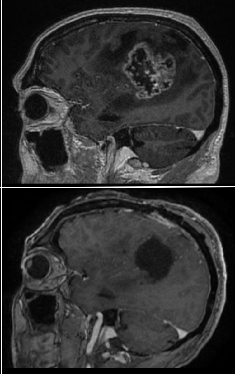
In a recent follow-up with a patient who was treated using this procedure, Dr. Valdes was happy to report that the individual was doing well nearly six months after having 100 percent of an enhancing tumor safely removed.
Comparisons of pre-and-post-op images of this patient, showcase the surgery's success, evidenced by a large void where the tumor mass once occupied.
Fluorescence-guided surgery is a common practice in other specialties, including breast and lung cancer cases, but its use in treating and removing brain tumors is newer and emerging.
“Certain agencies in the Texas Medical Center are not even doing it, so it’s pretty fantastic that we can do it here,” said Dr. Valdes of the leading-edge procedure.
For more information on Neurosurgery and other Neurosciences services, please visit the Neurosciences website.